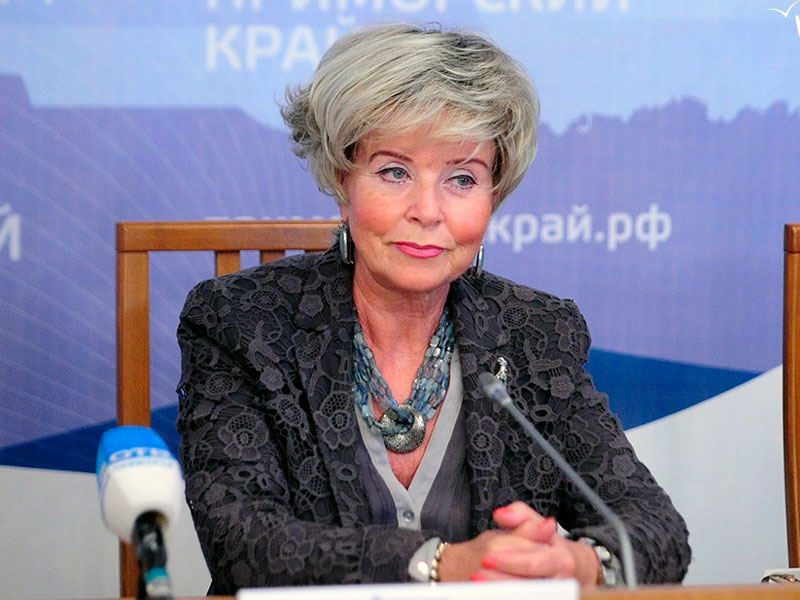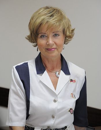
Interview on Breast Imaging Russia with Prof. Nadejda Rojkova
Breast imaging in Russia
An interview with Prof. Nadejda Rojkova, head of the Center of Oncology of Reproductive Systems
at Herzen Moscow Research Oncology Institute and Chief Specialist in mammology of the Russian Ministry of Health.
European Society of Radiology: Breast imaging is widely known for its role in the detection of breast cancer. Could you please briefly outline the advantages and disadvantages of the various modalities used in this regard?
Nadejda Rojkova: Various modalities of image acquisition are used to detect breast cancer: x-ray, ultrasound, MRI, scintigraphy, thermography, electrical impedance, optical (laser) imaging and others. The ‘gold standard’ is still breast mammography, which is used to diagnose most types of impalpable cancer. Among them, visible only on mammograms, are cancers in the form of assemblies of microcalcifications with dimensions of 50–400 microns (they make up 25% of cases of impalpable cancer), and cancers in the form of localised changes of breast tissue structure without the formation of a definite mass (another 17% of impalpable cancers). That is why mammography is widely used for screening. Other methods give additional information and are applied for more accurate diagnosis if needed. Thermography and electrical impedance mammography are sometimes used for young
women aged from 19 to 39 to define whether they are in the risk group.
ESR: Early detection of breast cancer is the most important issue for reducing mortality, which is one reason for large-scale screening programmes. What kind of programmes are in place in your country and where do you see the advantages and possible disadvantages?
NR: According to a special order issued by the Russian Ministry of Health (No.154 dated 2006), a
mammography procedure is applicable for all women older than 40 years. During the last ten years, up to the present moment, up to 68.5% of breast cancers have been detected in the early (1st and 2nd) stages. The one-year mortality rate from this disease has decreased to 26%. But insufficient medical coverage of the female population in huge remote territories of Russia makes the realisation of screening programmes quite difficult. Mass media, TV, public actions (‘open days’ at the screening centres), direct communication with women and their employers, are all used to increase the participation of female patients in screening programmes. During recent years the number of
asymptomatic women with silent forms of breast cancer detected at annual check-ups has increased up to 33%.
ESR: Do you know how many women take part (percentage) in Russian screening programmes? Do
patients have to pay for this?
NR: Mammography screening is free for patients and it is reimbursed through our state system of
obligatory medical insurance. Some forms of more detailed diagnostics have to be paid for. There are programmes for oncoepidemiological testing which are carried out for young women aged 19 to 39. If risk factors are detected, special training and consultations are given at health schools.
ESR: The most common method for breast examination is mammography. When detecting a possible
malignancy, which steps are taken next? Are other modalities used for confirmation?
NR: In order to detect breast cancer suspected at mammography, we perform biopsies followed by
cytological, histological and immunohistochemical examinations. We are looking for tissue biomarkers which could influence the selection of an appropriate treatment and definition of prognosis. This is a collaboration between a radiologist who does breast biopsies, a surgeon and a pathologist.
ESR: Diagnosing disease might be the best-known use of imaging, but how can imaging be employed
in other stages of breast disease management?
NR: Besides diagnosis, we use imaging to evaluate the effectiveness of treatment.
ESR: What should patients keep in mind before undergoing an imaging exam? Do patients undergoing radiological exams generally experience any discomfort?
NR: There is some radiation exposure during the mammography procedure. The patient is warned
about it and must sign an informed consent form before the procedure.
ESR: How do radiologists’ interpretations help in reaching a diagnosis? What kind of safeguards help to avoid mistakes in image interpretation and ensure consistency?
NR: The doctor takes into account pathomorphological characteristics of the lesion, not only
radiological ones. After that, an International Classification of Diseases (ICD) code about the type of disease is indicated in the report. The best medical schools train doctors in such imaging technologies as mammography, ultrasound and breast interventions, which helps them to make a diagnosis. Specialists in ultrasound and MRI prefer to use the BI-RADS classification system, though it is not used officially.
ESR: When detecting a malignancy, how is the patient usually informed and by whom?
NR: Oncologists inform patients about their diagnosis. They are also the ones who define further
management of the patient.
ESR: Some imaging technology, such as x-ray and CT, uses ionising radiation. How do the risks
associated with radiation exposure compare with the benefits? How can patient safety be ensured when using these modalities?
NR: Multiple trials have proven that the risk/benefit ratio for screening mammography is about
1:1,000. That is why mammography is a leading imaging modality used for screening and follow-up. Radiological safety measures such as protection by ensuring a suitable time and buffer distance, daily maintenance of equipment, and apron and throat protectors for the patient, are applied.
ESR: How aware are patients of the risks of radiation exposure? How do you address the issue with them?
NR: Patients sign an informed consent form.
ESR: How much interaction do you usually have with your patients? Could this be improved and, if
yes, how?
NR: According to order of the Ministry of Health, a 20-minute time slot is allocated for each patient coming for a breast check-up. It is used for communication with the patient, collection of relevant
medical information, physical examination and interpretation of x-ray images. If the doctor has an assistant, the time of the session can be further shortened.
ESR: How do you think breast imaging will evolve over the next decade and how will this change
patient care? How involved are radiologists in these developments and what other physicians are involved in the process?
NR: Radiology is booming. Nuclear medicine, non-ionising diagnostic imaging and telemedicine will be more widely used. Systems for teleradiology and teleconsultations have been created in order to improve the quality of diagnosis, decrease the need for qualified specialists, shorten the examination time for the patient, and speed up the start of treatment. In general, the process will become more efficient both diagnostically and economically.
In order to increase the accuracy of pre-operational diagnosis, interdisciplinary integration will be used together with biomolecular technologies. Thanks to such an approach, in complicated diagnostic cases, such biomarkers as the degree of cell apoptosis, proliferation, and others, will help in the detection and optimal treatment of impalpable breast tumours. Detection of the disease at the earliest stages and targeted diagnostics will contribute to the development of organ-preservation treatment technologies, aesthetic surgery, lipofilling and other methods to improve the cosmetic appearance after surgery. Development of technologies for cancer prevention will allow morbidity to be reduced further.
 Nadejda Rojkova, MD, PhD is a professor and Head of the Center of Oncology of Reproductive System at Herzen Moscow Research Oncology Institute in Moscow, Russia. She is also a Chief Specialist in mammology of the Russian Ministry of Health. She is a Chair of Clinical Mammology and serves as President of the Russian Association of Mammologists. She is an active member of many public organisations and a participant in various beneficial actions for the promotion of breast screening and women’s healthcare. She is an author and co-author of multiple scientific papers and more than 30 books on breast cancer imaging and treatment. Prof. Rojkova is a well-known speaker and teacher. In 2011–2015 she was President of the Russian Association of Radiologists. In 2014 she chaired the ‘ESR meets Russia’ session at the European Congress of Radiology in Vienna.